MedComm | Development of pathological reconstructed high-resolution images using artificial intelligence based on whole slide image
2020-12-17

Open the phone and scan
Pathological analysis is an important approach to diagnose diseases, particularly malignant tumors. The whole slide image (WSI), which meets the desired characteristics of high-definition, high-speed, and high-throughput screening, provides the possibility for the digitization of traditional pathology slices, which lays a solid foundation for the development and application of digital pathology (DP). With the development of DP, the storage and transmission of pathological images is now easier, which has led to many improvements in paleopathological consultation, digital management, and computer-assisted diagnosis by artificial intelligence (AI). There have been more than 1 000 000 cases of paleopathological consultation in China over the last 10 years.

In DP, the common digitization strategy is to scan the pathology slice with 20× or 40× objective. The data generated by the scan are called the WSI (or digital slide). Usually, the WSI of 40× is four times larger than that of 20× from the same slice, and hence, the storage space and transmission time of the data are four times. These increased costs are a significant negative factor in the popularization of DP. Although many lesions can be diagnosed at low or medium magnification, in some cases, even experienced pathologists need to observe cells and cell nuclear morphology at high magnification to further confirm the diagnosis. In this article, the authors present a novel reconstructed high-resolution (HR) process based on deep learning to switch 20 × WSI to 40 × without the loss of whole and local features. Furthermore, the authors collected the WSI data of 100 uterine leiomyosarcomas and 100 adult granulosa cell tumors to test the reconstructed HR process (Fig. 1). They tested the reconstructed HR WSI by the peak signal-to-noise ratio, structural similarity, and the blind/reject image spatial quality evaluator, which were 42.03, 0.99, and 49.22, respectively. Subsequently, they confirmed the consistency between the actual and reconstructed HR images. The testing results indicate that the reconstructed HR imaging is a reliable method for the digital slides of a variety.
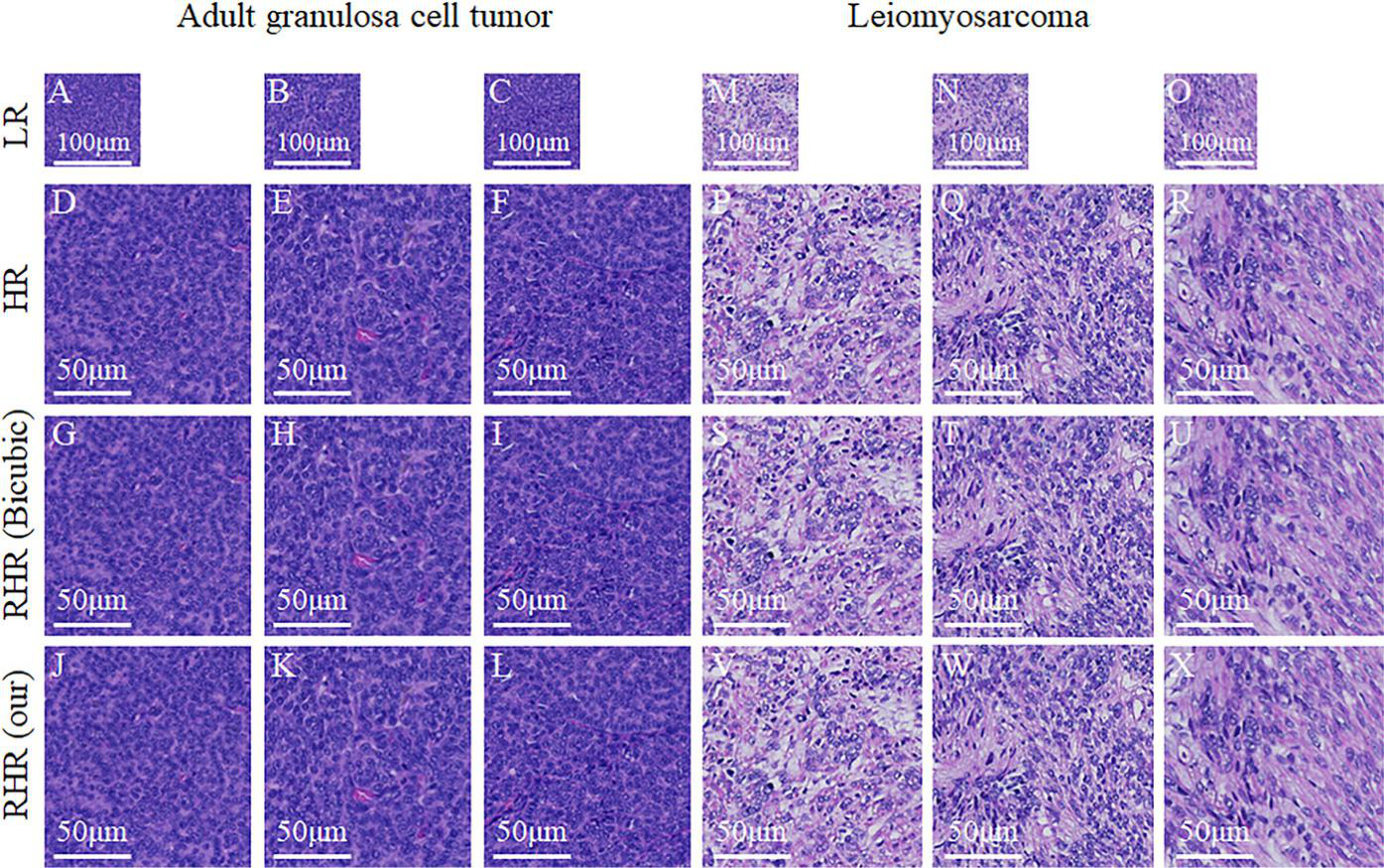
Fig.1 Result samples of AGCT and uterine leiomyosarcoma
Article Access: https://onlinelibrary.wiley.com/doi/10.1002/mco2.39
Website for MedComm: https://onlinelibrary.wiley.com/journal/26882663
Looking forward to your contributions.

